

Table of Contents
ToggleThree generations of cancer diagnostics. One blood draw that changed everything. And one question every oncologist and patient deserves an honest answer to.
Imagine you are trying to understand a city by taking a photograph of it.
- A tissue biopsy is like photographing one neighbourhood -one moment in time, one street corner, from one angle. It tells you something real and important. But it cannot tell you what the rest of the city looks like. It cannot tell you how the city is changing. And it definitely cannot tell you what the city will look like next year.
- Liquid biopsy zoomed out. Instead of one neighbourhood, it began sampling the entire city from above- detecting fragments of tumour DNA circulating in the bloodstream, gathered from multiple sites simultaneously. A significant leap.
- Cell biopsy – the third generation, goes further still. It doesn’t just photograph the city. It walks into a building, sits down with a resident, and asks them what’s happening inside, at the level of a single living cell.
This is not a metaphor. This is the actual trajectory of cancer diagnostics over the past three decades. And understanding where each approach sits, what it captures, what it misses, and when each one is the right tool- is one of the most clinically important conversations oncologists and patients can have today.
Generation 1: Tissue Biopsy:- The Gold Standard With Real Limitations
Tissue biopsy has been the cornerstone of cancer diagnostics for over a century – and it remains critically important. When a surgeon or interventional radiologist extracts a sample of tumour tissue for pathological examination, they are accessing the most direct source of information about a patient’s cancer.
From tissue, pathologists can assess tumour morphology, grade, receptor status, and; with an ngs test for cancer applied to the extracted DNA – a comprehensive landscape of the mutations driving the disease.
But tissue biopsy has three fundamental limitations that the field has known about for years and has been trying to solve ever since:
- Sampling bias. A biopsy captures one region of the tumour at one point in time. Tumours are not genetically uniform- they contain multiple subpopulations of cells with different mutations, different behaviours, and different responses to treatment. The region biopsied may not represent what is driving the most aggressive or resistant subclone. It’s like looking at only the trunk of the elephant and writing prescriptions based on the that. Missing a lot of important things on the other side.
- Invasiveness. Biopsies require procedures- needle insertion, bronchoscopy, endoscopy, or surgery, depending on tumour location. For deep-seated tumours or patients in poor condition, repeat biopsy at progression is simply not feasible. Yet the tumour’s biology at progression; when treatment resistance has emerged, is exactly when re-profiling is most clinically valuable.
- Static snapshot. Cancer evolves. The molecular profile at diagnosis is not the same as the profile six months into treatment. Tissue biopsy cannot track this evolution without repeat procedures; and repeated invasive biopsies are neither practical nor always safe.
It’s painful at the end of the day.
These are not criticisms of tissue biopsy. They are realities of the biology it is trying to capture, and they are exactly what the second and third generations of cancer diagnostics were built to address.
Generation 2: Liquid Biopsy and the ctdna test- A Leap Forward
The concept behind liquid biopsy is elegantly simple: tumours shed DNA into the bloodstream as cells die and break down. This circulating tumour DNA- ctDNA; can be extracted from a blood sample and analysed using the same comprehensive genomic profiling techniques applied to tissue.
The implications were immediately transformative:
- A simple blood draw replaces an invasive procedure
- The blood samples signals from multiple tumour sites simultaneously — not just the biopsied region
- The test can be repeated as often as clinically needed — tracking how the tumour evolves in response to treatment
- A ctdna test can detect emerging resistance mutations in real time, before they cause clinical or radiological progression
For patients and oncologists, the liquid biopsy test india market has grown rapidly; making non-invasive tumour profiling increasingly accessible across major oncology centres. The ability to monitor disease without repeat biopsies is genuinely life-changing for patients who have been through surgery, chemotherapy, and radiation and face years of follow-up.
In MRD monitoring specifically, the ctdna test has been transformative. Studies show ctDNA can detect breast cancer recurrence up to 18.5 months before clinical or radiological evidence appears; and the NCCN now formally recommends ctDNA-based minimal residual disease testing in bladder cancer, with evidence building across multiple tumour types.
But ctDNA-based liquid biopsy has its own ceiling; and the field is now confronting it honestly.
The limits of ctDNA:
- At very low tumour fractions; particularly in early-stage disease or in the post-treatment setting where very few tumour cells remain – ctDNA sensitivity drops significantly
- ctDNA captures only what tumour cells have shed into the bloodstream as they die. It cannot capture the biology of live, circulating tumour cells; the cells that are actively evading the immune system, travelling to distant sites, and seeding metastases
- ctDNA is DNA only. It cannot tell you what genes are switched on (RNA), or what proteins are expressed on the cell surface (proteomics); the layer that matters most for immunotherapy selection and ADC target identification
- In our own ASCO 2026 research on metastatic pancreatic cancer, 65% of genomic alterations were found exclusively in single-cell CTC-DNA; completely absent from matched ctDNA. The tumour biology was there. The ctDNA just couldn’t see it
This is the ceiling of generation two. And it is what makes generation three- Cell Biopsy® – such a significant clinical leap.
Generation 3: Cell Biopsy® – The Complete tumor profiling test
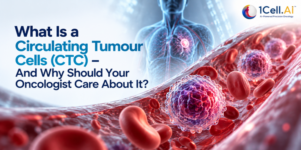
Cell Biopsy® is not an incremental improvement on liquid biopsy. It is a fundamentally different kind of information; because it analyses live, individual Circulating Tumour Cells (CTCs) rather than fragments of DNA left behind by dead ones.
When tumours progress, metastasize, or develop resistance to treatment, they shed entire living cells into the bloodstream. These CTCs carry the full molecular complexity of the tumour; not just their DNA, but their RNA (what genes are active right now), their protein expression (what is sitting on the cell surface, targetable by therapy), and their epigenetic state. Capturing and analysing these cells, at the resolution of a single cell, delivers a view of tumour biology that neither tissue biopsy nor ctDNA liquid biopsy can match.
At 1Cell.Ai, our OncoIncytes™ platform – the clinical implementation of Cell Biopsy®, achieves CTC isolation at a sensitivity of 1 in a billion cells, capturing live CTCs from just 10-20 mL of blood. Crucially, we isolate CTCs without fixing them; preserving the cell’s natural state and enabling true downstream multi-omics analysis.
From a single blood draw, OncoIncytes™ delivers:
- DNA: identifying mutations, fusions, copy number variants, HRD, TMB, MSI across our OncoIndx® 1,080-gene panel, with comprehensive genomic profiling at 10,000x sequencing depth for ctDNA
- RNA: whole transcriptome sequencing revealing gene expression, novel fusions, splice variants, and pathway activation patterns invisible to DNA alone
- Protein: direct measurement of cell surface protein expression (PD-L1, HER2, TROP-2, Claudin 18.2, and more), the layer that determines ADC eligibility and immunotherapy response
No other liquid biopsy platform currently combines all three simultaneously from live single cells.
Head-to-Head: What Each Generation Captures
| Tissue Biopsy | Liquid Biopsy (ctDNA) | Cell Biopsy® (CTC) | |
| Sample | Tumour tissue | Blood
|
Blood
|
| Invasiveness | Invasive procedure | Blood draw
|
Blood draw
|
| DNA mutations | High depth | Limited by tumour fraction | Single-cell resolution |
| RNA / gene expression | ✅ Possible from tissue | ❌ Not available | ✅ Single-cell transcriptomics |
| Protein expression | ✅ IHC from tissue
|
❌ Not available
|
✅ Direct cell surface profiling |
| Tumour heterogeneity | Single region only | Bulk signal: averages subclones | ✅ Individual cell-level resolution |
| Real-time monitoring
|
❌ Repeat biopsy needed | ✅Repeatable blood draws | ✅ Repeatable blood draws |
| MRD sensitivity | ❌ Not applicable | ✅ High at sufficient tumour fraction | ✅ Detects viable circulating cells |
| ADC target identification | Static tissue snapshot | ❌ Not available | ✅ Live protein expression on CTCs |
| Treatment resistance | Requires re-biopsy | Detects ctDNA resistance mutations | Captures live resistant subclones
|
| Tumour of origin (CUP) | ✅ Morphology-based | Limited | ✅ Multi-omic molecular similarity |
When to Use Each? – A readymade framework for clinicians
These three generations are not mutually exclusive. They answer different clinical questions at different points in a patient’s journey, and the most comprehensive approach integrates all three where feasible.
Use tissue biopsy when:
- Initial diagnosis and histological classification is needed
- Tumour-specific morphology assessment is required
- Tissue is accessible and the procedure is safe
Use a ctdna test (liquid biopsy) when:
- Repeat profiling at progression or resistance is needed without re-biopsy
- Post-surgical minimal residual disease testing is the clinical question
- Monitoring treatment response longitudinally
- Tissue is inaccessible or patient is not fit for procedure
Use Cell Biopsy® (OncoIncytes™) when:
- A driver-negative ctDNA result leaves the treatment question unanswered
- ADC therapy selection requires protein expression data from live tumour cells
- Tumour heterogeneity at the single-cell level is the clinical priority
- The patient needs the most comprehensive tumor profiling test available – DNA + RNA + protein from a single blood draw
For the highest-complexity cases; advanced disease, exhausted options, unknown primary, or early-stage clinical trial enrolment- integrating ctDNA and CTC analysis together delivers a panoramic view of the tumour that neither modality provides alone.
The Future of tumor profiling test: Integration, Not Replacement
The history of cancer diagnostics is not one technology replacing another. And it’s definitely not a competition. It is one technology adding to the last; each generation closing the gaps the previous one left open.
It’s a group project, not a race.
Tissue biopsy gave us the histological foundation. The liquid biopsy test india market demonstrated that blood could tell a meaningful tumour story non-invasively. And Cell Biopsy® is now proving that live, single-cell multi-omics delivers a layer of tumour intelligence that neither of its predecessors could access.
For precision oncology india- a market where over 70% of cancers are diagnosed at advanced stages, where repeat tissue biopsies are often not feasible, and where the most vulnerable patients need the most powerful tools; this progression is not academic.
It is clinical necessity.
At 1Cell.Ai, OncoIncytes™ and Cell Biopsy® represent our answer to the question every oncologist is increasingly asking:
“Is there a more complete version of this information?”
There is.
It starts with a blood draw. And it ends with a molecular portrait of the tumour that no single generation of diagnostics could have produced alone.
To learn more about OncoIncytes™ and our Cell Biopsy® platform, visit 1cell.ai or reach us at [email protected]