

Table of Contents
ToggleTumour Heterogeneity – No Two Cancer Cells Are the Same
Tumors do not evolve as homogenous tissue masses, but rather as dynamic, branch-evolving ecosystems characterized by extensive intra-tumor heterogeneity (ITH). Within a single oncology patient, distinct subclonal populations coexist, harboring divergent somatic copy-number alterations (SCNAs), single-nucleotide variants (SNVs), and transcriptional states.
This internal diversity is the main enemy in our fight against cancer. The diversity within tumor cells demands diversity in our treatment approaches, but unfortunately, that doesn’t exist yet.
While a primary frontline therapy may successfully destroy 99% of a tumor mass, rare subclonal cells (such as those undergoing epithelial-to-mesenchymal transition, or EMT) often carry intrinsic resistance mechanisms. These cells survive the initial therapeutic bottleneck, driving disease recurrence and distal metastasis.
┌──► Subclone A (Proliferative) ──► Sensitive to Platinum Chemotherapy
│
Tumor ─┼──► Subclone B (Dormant Stem) ──► evades cytotoxic agents, drives recurrence
│
└──► Subclone C (EMT Phenotype) ──► Enters circulation as CTCs, drives metastasis
What Bulk Testing Misses
Standard clinical molecular diagnostics rely on bulk Next-Generation Sequencing (NGS). Bulk sequencing extracts genomic material from a blended tissue or plasma sample, generating an arithmetic mean of all cellular signals present.
Rare resistant subclones often remain below the detection threshold of conventional bulk assays, masking the true biological complexity of the tumor.
| Feature | Bulk NGS | Single cell analysis |
| Unit of analysis | Millions of cells combined | One cell at a time |
| Detects rare resistant clones | Often missed | Easily identifiable |
| Tumor heterogeneity | Averaged signal | Full cellular diversity |
| Clonal evolution tracking | Limited | High resolution |
| Therapy resistance detection | Often delayed | Early detection |
| Precision oncology value | Moderate | Very high |
The Single-Cell Revolution
The single-cell revolution resolves this blind spot by isolating individual cellular entities prior to amplification and sequencing. This shifts our diagnostic capability from an aggregate overview to individual, single-cell resolution. It allows clinicians to map the absolute clonal hierarchy of a malignancy, capture highly elusive stem-like cancer cells, and detect emergent resistance patterns before they manifest as physical macro-metastases on radiological scans.
What is Multi-Omics?
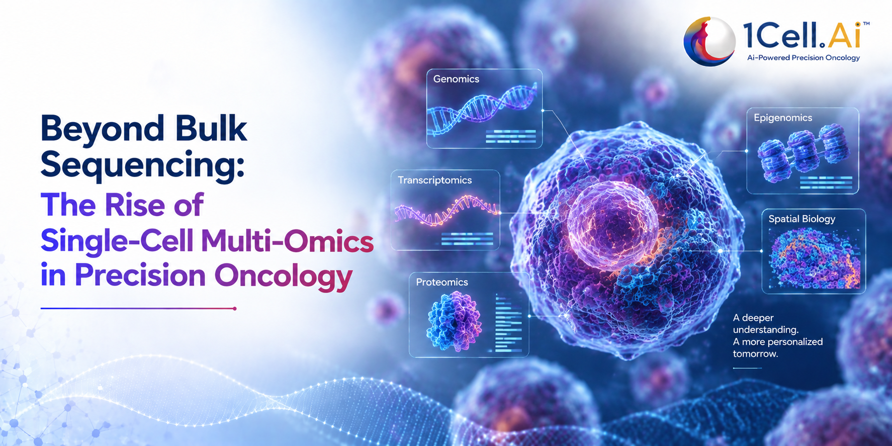
Genomics, Transcriptomics, Proteomics
True single-cell multi-omics requires the simultaneous extraction and analysis of multiple molecular layers from a single, undivided physical cell:
- Genomics (DNA): Defines the hardwired structural blueprint, documenting somatic mutations, insertion-deletions (indels), copy-number variations (CNVs), and passenger vs. driver alterations.
- Transcriptomics (RNA): Measures the functional output of the genome through messenger RNA (mRNA) expression, revealing which pathways (e.g., MAPK, PI3K/Akt) are actively upregulated.
- Proteomics (Proteins): Captures the final phenotypic reality via functional cell-surface markers and intracellular signaling proteins, which dictate immune interactions and direct drug binding.
Why Analysing One Layer at a Time Is Not Enough
Evaluating a single omic layer in isolation generates dangerous clinical assumptions. Intracellular information transmission is non-linear and heavily modified by epigenetic factors, post-transcriptional silencing, and translational regulation.
For instance, a cell may harbor an EGFR gene amplification (Genomic layer). However, if that gene is epigenetically silenced or fails to properly translate into active EGFR membrane receptors (Proteomic layer), targeting that patient with an EGFR tyrosine kinase inhibitor (TKI) will yield zero therapeutic response.
The Power of Integration- Seeing the Full Picture
By performing integrated single-cell multi-omics, we map the direct, causal flow of biological information across layers within the exact same cell. This allows investigators to definitively correlate genetic driver mutations directly with corresponding transcriptional activity and real-time protein biomarker density (e.g., co-localizing HER2 copy-number gains with actual surface HER2 protein overexpression).
Single-Cell Multi-Omics in Practice
How Single-Cell Analysis Works
Single-cell multi-omics follows a highly structured workflow from cell isolation to computational interpretation.
Blood Sample → Single Cell Isolation → Molecular Barcoding → High-Depth Sequencing → Bioinformatics Analysis → Clinical Insights
- Microfluidic Compartmentalization: Individual cells are isolated into separate compartments.
- Multimodal Barcoding: Unique molecular barcodes label DNA, RNA, and proteins from each cell.
- High-Depth Next-Generation Sequencing (NGS): Barcoded molecules are sequenced simultaneously.
- Bioinformatics analysis: Computational pipelines reconstruct the molecular profile of every cell.
What DNA, RNA, and Protein Data From One Cell Can Tell Us
Simultaneous multi-layered single-cell reads expose complex phenotypes like Homologous Recombination Deficiency (HRD), matching structural genomic scars directly to underlying transcription changes and immune escape markers like PD-L1. This resolution exposes whether an aggressive subclone is actively deploying protective immune-checkpoint mechanisms or shifting its cellular state to bypass current therapies.
Applications in Drug Discovery and Clinical Trials
For biopharma translational workflows, single-cell multi-omics streamlines the drug development pipeline:
Target Identification: Uncovering rare, highly specific cell-surface targets restricted to malignant lineages while sparing healthy tissue.
- Mechanism of Action (MoA) Mapping: Directly observing how therapeutic candidates alter downstream transcriptomic networks and protein expressions across heterogeneous cell populations.
Clonal Evolution Tracking: Longitudinally monitoring clinical trial cohorts to map the precise molecular pathways cells utilize to develop acquired drug resistance, enabling the rational design of combination therapies.
Circulating Tumour Cells- The Ideal Single-Cell Source
What Are CTCs and Why Are They Special?
To fully utilize single-cell multi-omics over a patient’s care timeline, clinicians require a repeatable, non-invasive source of tumor cells. Serial solid-tissue biopsies are highly invasive, carry surgical risks, and are prone to severe spatial sampling bias.
Circulating Tumor Cells (CTCs) are intact, living malignant cells shed from primary or metastatic sites that enter the peripheral circulation. Unlike ctDNA, CTCs preserve intact cellular architecture, making them ideal for single-cell multi-omic analysis. They preserve the complete, un-fragmented spatial architecture of the tumor’s DNA, RNA, and proteins, serving as an easily accessible, real-time “live tissue biopsy.”
| Feature | CTCs | ctDNA |
| Cell status | Living intact cells | Fragmented DNA |
| DNA analysis | ✅ | ✅ |
| RNA analysis | ✅ | ❌ |
| Protein analysis | ✅ | ❌ |
| Functional studies | ✅ | ❌ |
| Single cell sequencing | ✅ | ❌ |
| Longitudinal monitoring | ✅ | ✅ |
The Challenge of Isolating Live CTCs
Despite their immense diagnostic value, isolating CTCs has been a historical bottleneck. Standard isolation methods often rely on chemical fixation, which destroys delicate mRNA networks and distorts real-time protein expression. Furthermore, aggressive mechanical sorting platforms expose cells to high shear stresses, killing the cells or triggering artifactual stress-response pathways that mask their true biological state.
One in a Billion- The Sensitivity That Makes It Possible:-
The primary challenge is an extreme needle-in-a-haystack problem. In advanced-stage cancer patients, a standard 10 mL blood sample contains approximately 50 billion red blood cells and 50 million white blood cells (leukocytes). Interspersed among them are often fewer than 1 to 10 CTCs. Achieving clinical utility requires an isolation platform with an analytical sensitivity capable of identifying a single malignant cell out of billions of background blood cells, while ensuring zero leukocyte contamination to protect downstream multi-omic assays.
How 1Cell.ai is Leading Single-Cell Multi-Omics
Our Cell Biopsy® Technology- Capture, Release, Analyse
1Cell.ai has overcome these isolation challenges by building a proprietary, clinical-grade precision oncology platform around its flagship Cell Biopsy® technology. Moving beyond the constraints of dead ctDNA fragments, the platform captures, releases, and profiles fully intact, live CTCs through a tightly orchestrated process:
Capture: Utilizing advanced microfluidic chips engineered with highly selective, affinity-based surface chemistry, the platform captures rare, viable CTCs directly from peripheral whole blood with high efficiency, while allowing background blood components to pass through unharmed.
Release: The platform applies a proprietary, ultra-gentle enzymatic and fluidic release mechanism that lifts the bound CTCs cleanly off the microfluidic substrate. This process avoids chemical fixation and high shear forces, keeping the isolated tumor cells live, intact, and biologically un-manipulated.
Analyse: The freed single cells flow directly into the automated 1Cell SOLO® system. This system utilizes AI-driven visual selection and advanced microfluidics to isolate true single cells, preparing them for deep, high-purity single-cell genomic, transcriptomic, and proteomic multi-omic sequencing workflows.
From Blood Draw to Multi-Dimensional Tumour Intelligence
Once sequencing is complete, the raw data enters the 1Cell iCORE® platform, an advanced AI-powered informatics and cloud infrastructure engine. The iCORE system synthesizes the multi-layered molecular datasets, separating true tumor signals from background noise, tracking clonal evolution over time, and generating an integrated, actionable report aligned directly with ASCO, ESMO, and NCCN clinical guidelines.
Real-World Applications- PDAC, NSCLC, Breast Cancer and Beyond:
The clinical utility of the 1Cell.ai ecosystem has been validated across more than 40 peer-reviewed abstracts and poster presentations at major international oncology conferences, including the American Society of Clinical Oncology (ASCO) and the American Association for Cancer Research (AACR):
1. Pancreatic Ductal Adenocarcinoma (PDAC): Pancreatic tumors are notoriously hard to tissue-biopsy and exhibit extreme internal heterogeneity. Data presented at ASCO demonstrated the platform’s ability to track single-CTC clonal evolution over time, mapping co-occurring KRAS and TP53 mutations within rare, highly aggressive subclonal populations driving therapy resistance.
2. Non-Small Cell Lung Cancer (NSCLC): Measuring PD-L1 expression via traditional tissue immunohistochemistry (IHC) frequently misses structural variations due to localized tissue sampling errors. 1Cell.ai platforms successfully map out single-CTC surface PD-L1 protein expressions in tandem with underlying genomic rearrangements (such as EGFR mutations), delivering an accurate, longitudinal predictor of immunotherapy response directly from a blood draw.
3. Breast Cancer: As patients progress through complex lines of targeted therapies, including Selective Oestrogen Receptor Degraders (SERDs) and Antibody-Drug Conjugates (ADCs), the 1Cell.ai platform continuously monitors shifting surface protein levels (e.g., HER2 and PD-L1 alterations) alongside emerging resistance mutations (e.g., ESR1 and PIK3CA variants), catching therapeutic escape pathways weeks before they appear on standard radiological scans.
What This Means for the Future of Cancer Care?
Personalised Therapy Selection at Single-Cell Resolution
The clinical integration of 1Cell.ai’s single-cell multi-omics marks a major shift from reactive treatment to proactive precision oncology. By resolving the exact molecular mechanisms inside the most resistant single cells, clinicians can move past treatment choices based on simple tissue averages, selecting highly targeted therapies tailored to eradicate the specific subclonal drivers of a patient’s unique disease.
Accelerating the Next Generation of Cancer Drugs
For global pharmaceutical and translational research organizations, this integrated platform accelerates clinical trial timelines. By providing clear, longitudinal evidence of patient response, biomarker expression, and emergent resistance mechanics at single-cell resolution, the platform significantly de-risks drug development and empowers the design of highly effective, next-generation combination therapies.
Conclusion
The historic paradigm of treating cancer through bulk averages or fragmented ctDNA footprints is being replaced by single-cell resolution. Through its Cell Biopsy® isolation technology, 1Cell SOLO® hardware, and iCORE® AI informatics engine, 1Cell.ai is transforming multi-dimensional single-cell multi-omics into an accessible, real-time clinical reality, unlocking cell-level intelligence to reshape the future of precision oncology.