


The treatment was working.
The scans were improving. The tumor markers were settling. For the first time in months, there was room to breathe.
And then a follow-up scan showed something no one wanted to see — the disease was growing again.
It is one of the hardest moments in oncology, for the patient and the physician alike. A therapy that was clearly working simply stops working. The natural question is short, but the answer rarely is:
Why did the treatment stop working?
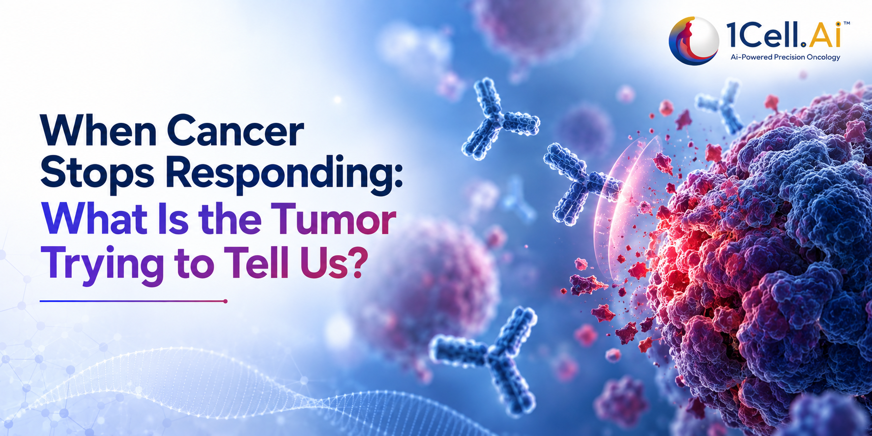
The reason is that cancer is not a fixed target. A tumor is a mixed population of cells, and treatment does not act on all of them equally. Some cells die. A few survive. And the ones that survive may carry exactly the traits that let them keep growing while the therapy is still being given.
We call this treatment resistance and understanding it has quietly become one of the most important questions in modern cancer care. The question is no longer only “what mutation does this cancer have?” It is also “what is this cancer becoming?”
What do we actually mean by “resistance”?
Resistance simply means the cancer no longer responds to a therapy either from the very start, or after an initial period of benefit.
When it is present from the beginning, we call it primary (or intrinsic) resistance: the treatment never really worked. When a cancer responds well and then begins to grow again, that is acquired resistance: the tumor learned to escape.
The distinction matters, because it changes what we do next.
It also reminds us of something easy to forget. The molecular profile taken at diagnosis is a snapshot – an accurate picture of the cancer at that moment. Under the pressure of treatment, some cell populations shrink, others expand, and new molecular changes can surface.
A tumor is not a photograph. It is a film that keeps rolling long after the first frame.
Why does cancer become resistant?
It helps to think of a tumor as a population rather than a single, uniform mass. Different cancer cells within the same tumor can carry different genetic alterations and behaviors – a property called tumor heterogeneity.
When treatment begins, the sensitive cells are cleared first. But a small resistant subpopulation may survive, multiply, and gradually take over. Over time, the tumor effectively rewrites itself. This is clonal evolution wherein cancer is adapting, generation by generation, to the pressure we apply.
That adaptation can take several forms:
- A change in the exact gene or protein the drug targets
- Activation of an alternative signaling route that bypasses the block
- Amplification or loss of a gene
- Shifts in gene expression or epigenetic state
- Changes in the tumor’s surrounding microenvironment
The examples are well documented. In EGFR-mutant lung cancer, tumors controlled by a targeted inhibitor can return with a new alteration such as EGFR C797S or MET amplification that reopens the growth signal. In hormone-receptor-positive breast cancer, ESR1 mutations can emerge during endocrine therapy and drive resistance that was not present at diagnosis. In many cancers, tumors reactivate the MAPK or PI3K/AKT pathways to find a way around a blocked target.
So resistance is rarely a single event.
Resistance is not always one new mutation. It is often several escape routes opening at once.
The biopsy from diagnosis may not describe today’s tumor
Picture the usual sequence. A patient is diagnosed, a biopsy is taken, the tumor is profiled, an actionable alteration is found, and treatment is chosen accordingly.
Months later, the cancer progresses.
One likely explanation is simply that the tumor has moved on. The cancer being treated today may no longer be biologically identical to the cancer that was profiled at diagnosis.
Which raises a fair clinical question: should decisions at progression still rest entirely on the original molecular report?
Not always. In selected patients with progression or suspected acquired resistance, taking a fresh look at the tumor’s biology can reveal why it escaped and sometimes point to what to do next.
Progression is not always the end of options. Sometimes it is the tumor handing us a new clue.
From a snapshot to a moving picture
Conventional testing tends to work in still images: a biopsy here, a scan there, a blood test in between. But the disease between those images is continuously changing.
This is where longitudinal molecular monitoring which follows the tumor over time rather than at a single point becomes genuinely useful. Instead of asking only “what mutations are present?”, we can ask “how is this molecular picture shifting?”
One practical way to follow those shifts is a liquid biopsy, a blood test, that looks for tumor signals rather than requiring another tissue sample. Much of it centers on circulating tumor DNA (ctDNA) which are fragments of DNA shed by cancer cells into the bloodstream. Because blood can be drawn repeatedly, ctDNA offers a way to watch the molecular story evolve during treatment.
Liquid biopsy does not replace tissue biopsy in every case. But because it is minimally invasive, it makes repeated assessment realistic in the right clinical setting and repetition is exactly what tumor evolution demands.
There is an important caveat, though. A negative liquid biopsy does not prove resistance is absent. Tumors differ in how much ctDNA they release, and disease burden, location and biology all affect what can be detected. Molecular results are a piece of the picture, never the whole of it and must always be read alongside imaging, pathology and the patient’s clinical course.
What if the resistance isn’t in the DNA?
Genomics is powerful, but it does not explain everything.
Cancer cells can survive treatment by changing which genes they switch on, by altering their cellular state, or by leaning on their surroundings all without an obvious new DNA mutation. A tumor can look genomically “quiet” and still be actively escaping.
That is why precision oncology is increasingly reaching beyond DNA alone. RNA shows which genes are actually active. Protein-level analysis shows which pathways are truly switched on. And single-cell approaches can expose differences between individual tumor cells that a whole-tumor average would blur away.
The aim is not simply to generate more data.
When DNA falls silent, the answer may be written in RNA or protein instead.
What this means in the clinic
None of this argues for re-testing every patient at every turn. Repeat molecular testing is a considered decision shaped by the cancer type, therapy, clinical course, quality of the original sample, the resistance mechanism suspected, and whether a result would actually change management.
But when a cancer progresses, one question is always worth asking clearly: do we understand why?
Depending on the situation, that answer may come from a repeat tissue biopsy, a liquid biopsy, comprehensive genomic profiling, RNA-based testing, or additional biomarkers and most often from thoughtfully combining several of these with imaging and clinical judgement. The most informative approach is rarely a single test in isolation.
How 1Cell.Ai approaches the moving target
If resistance is a moving target, testing has to move with it and that is where a longitudinal, multi-modal approach earns its place.
For patients on treatment, OncoAlibrex®, 1Cell.Ai’s treatment-response monitoring solution, is designed to follow the disease over time using serial liquid-biopsy signals, so that a molecular shift can be noticed and discussed rather than waiting for the next scan to declare it.
When the question is deeper - why has this tumor escaped, and is the reason even in the DNA? — OncoIndx® Multi-Modal brings together DNA, RNA and protein in a single integrated profile, precisely because resistance is not always a genomic event. Where a broad genomic re-assessment at progression is the priority, OncoIndx® Prime+ provides comprehensive genomic profiling across tissue and liquid biopsy.
All of this sits on 1Cell.Ai’s OncoIncytes® platform, which analyses ctDNA together with circulating tumor cells at single-cell resolution alongside transcriptomic and proteomic data in an attempt to see not just what the tumor is, but what it is doing.
The point of these tools is not more testing for its own sake. It is to keep the molecular picture current, so that each decision is made against the cancer as it exists now and not the cancer as it existed at diagnosis.
The takeaway
For a long time, cancer care asked one question: what cancer is this? Precision oncology added a second: what mutation does it carry? We are now learning to ask a third, and perhaps the most important one: how is this cancer changing?
If a tumor progresses after responding, it does not necessarily mean the options have run out. Often it means the biology has shifted and that shift, once understood, can guide what comes next. Those conversations belong with the treating oncology team, but understanding the science behind resistance helps everyone in the room ask better questions.
The most useful test is not always the first one you did. Often it is the one that tells you what has changed.
Because in the end, precision oncology is not a single decision made once. It is a series of decisions, each one kept honest to the cancer as it exists today.